Understanding the Differences Between Types of Medical Imaging
Medical imaging is a key tool in modern medicine, helping doctors see inside your body in detail. Common types of imaging include:

- X-rays
- CT scans (computed tomography)
- MRI (magnetic resonance imaging)
- Ultrasounds
- PET scans (positron emission tomography)
Each imaging technique uses a different technology and is suited for specific purposes. Medical imaging technologies are mostly used for medical diagnoses. For example, X-rays are great for spotting bone fractures, while MRIs are better for examining ligament injuries.
X-ray
X-rays are quick, painless tests that produce images of the structures inside your body, especially bones.
What to expect during an X-ray
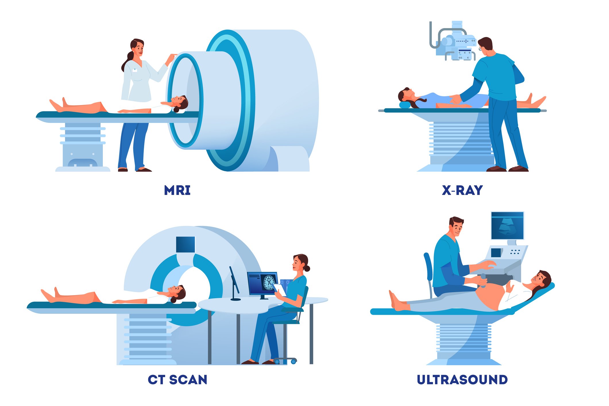
You will lie, sit, or stand while the X-ray machine takes images. One piece of equipment sends the radiation out and another contains the X-ray film. You may be asked to move in several positions.
- Duration: 10-15 minutes
- Imaging Method: X-rays use ionizing radiation. The amount of radiation is relatively low but can accumulate over multiple exposures.
- Used to diagnose:
- Bone fractures
- Arthritis
- Osteoporosis
- Infections
- Breast cancer
- Swallowed items
- Digestive tract problems
- Possible Risks: Exposure to ionizing radiation can increase the risk of cancer over time, although the risk from a single X-ray is minimal.
Computed Tomography (CT) Scans
CT scans use a series of X-rays to create cross-sections of the body, including bones, blood vessels, and soft tissues.
What to expect during a CT scan
You will lie on a table that slides into the scanner, which looks like a large doughnut. The X-ray tube rotates around you to take images.
- Duration: 10-15 minutes
- Imaging method: CT scans involve a higher dose of ionizing radiation compared to standard X-rays, but they provide more detailed images.
- Used to diagnose:
- Injuries from trauma
- Bone fractures
- Tumors and cancers
- Vascular disease
- Heart disease
- Infections
- Used to guide biopsies
- Possible Risks: The increased radiation dose can pose a higher risk of cancer, especially with frequent scans.
Magnetic Resonance Imaging (MRI)
MRIs use magnetic fields and radio waves to create detailed images of organs and tissues in the body.
What to expect during an MRI
You will lie on a table that slides into the MRI machine, which is deeper and narrower than a CT scanner. The MRI magnets create loud tapping or thumping noises.
- Duration: 30-60 minutes
- Imaging method: MRI does not use ionizing radiation. Instead, it relies on magnetic fields and radio waves.
- Used to diagnose:
- Aneurysms
- Multiple sclerosis (MS)
- Stroke
- Spinal cord disorders
- Tumors
- Blood vessel issues
- Joint or tendon injuries
- Possible Risks: MRI is generally considered safe, but patients with certain metal implants or devices may face risks. Claustrophobia can also be a concern due to the machine's confined space.
Ultrasound
Ultrasound uses high-frequency sound waves to produce images of organs and structures within the body.
What to expect during an ultrasound
A technician applies gel to your skin, then presses a small probe against it, moving it to capture images of the inside of your body.
- Duration: 15-45 minutes
- Imaging method: Ultrasound does not use ionizing radiation. It relies on sound waves to produce images.
- Used to diagnose:
- Gallbladder disease
- Breast lumps
- Genital/prostate issues
- Joint inflammation
- Blood flow problems
- Monitoring pregnancy
- Used to guide biopsies
- Possible Risks: Ultrasound is generally very safe with no known risks related to ionizing radiation
Positron Emission Tomography (PET)
PET scans use radioactive drugs (called tracers) and a scanning machine to show how your tissues and organs function.
What to expect during a PET scan
You swallow or have a radiotracer injected. You then enter a PET scanner (which looks like a CT Scanner) which reads the radiation off by the radiotracer.
- Duration: 1.5-2 hours
- Imaging method: PET scans involve exposure to radioactive tracers emitting ionizing radiation, though the amount is typically low.
- Used to diagnose:
- Cancer
- Heart disease
- Coronary artery disease
- Alzheimer’s disease
- Seizures
- Epilepsy
- Parkinson’s disease
- Possible Risks: Exposure to radioactive tracers carries a small risk of cancer, and there may be reactions to the tracer in some individuals.
Each imaging technique offers unique benefits and is suited for specific diagnostic purposes. Understanding these differences helps patients make informed decisions about their healthcare. Always discuss with your healthcare provider to determine which imaging method is best suited for your condition and to understand any associated risks. Visit PIHHealth.org/Radiology to learn more about imaging at PIH Health.